

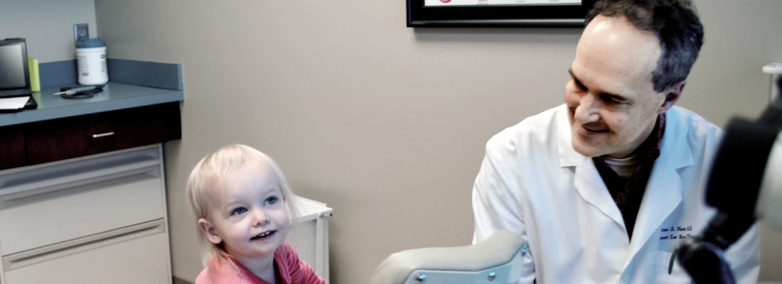


Earaches
Earache, or pain in the ear, is common and can occur in both children and adults. It can be due to a problem with the ear or structures close to the ear. The pain may be dull, sharp, or burning and can occur in one or both ears. It may be constant or come and go.
What Are the Symptoms of Earaches?
Symptoms of earaches in infants and toddlers may include:
- Hearing problems
- Pulling or scratching the ear
- Crying or irritability
- Ear drainage
- Fever
Symptoms of earaches in young children, adolescents, and adults may include:
- Pain
- Hearing problems
- Full or “stuffy” sensation in the ear
- Dizziness or loss of balance
- Nausea, vomiting
- Ear drainage
- Fever
What Causes Earaches?
A variety of problems can cause earaches in children and adults:
- Middle ear infection (called acute otitis media)
- Swimmer’s ear (called otitis externa)
- Temporomandibular joint (TMJ) dysfunction or jaw joint pain
- Eustachian tube dysfunction
- Inflammation of the outer ear (called chondritis)
- Cotton swab use
- Throat infection
- Throat cancer (rarely)
What Causes Earaches in Children?
In children, earaches are commonly due to an infection of the middle ear (acute otitis media), and can affect one or both ears. Otitis media can be serious because the infection can spread to nearby structures in the head, especially the mastoid located behind the ear. Otitis media may also cause hearing loss; in children, it may impair learning ability and even delay speech development. However, if it is treated promptly and effectively, hearing can almost always be restored to normal.
Many cases of otitis media can be treated by your pediatrician or family doctor; more serious cases may need attention from an ENT (ear, nose, and throat) specialist, or otolaryngologist.
Ear infections are often due to eustachian tube dysfunction. Inside your ear you have something called an eustachian tube that equalizes pressure behind the ear drum and naturally clears middle-ear secretions. When the eustachian tube becomes blocked due to a cold, allergy, upper respiratory infection (URI), bacteria, or a virus, negative pressure can develop and mucus can collect behind the eardrum causing pain, swelling, and redness.
What Causes Earaches in Adults?
In adults, common causes of earaches include otitis externa or swimmer’s ear and TMJ dysfunction. Swimmer’s ear is an infection of the ear canal and results from swimming in contaminated water or as a result of cotton swab use. In people with diabetes, otitis externa can spread far beyond the ear canal and can be life threatening. Therefore, prompt treatment with antibiotic ear drops as well as cleaning of the ear canal with specialized tools available to an ENT specialist is critical. Another common cause of ear pain is due to referred pain from the jaw joint. This is usually due to grinding of teeth during sleep. TMJ pain can be treated with ibuprofen, eating softer food, avoiding chewing gum, and using a night guard.
An avoidable cause of earache is the use of cotton swabs or other instruments to clean wax from the ear, which can damage the ear canal. Following the old adage that “nothing smaller than an elbow goes in the ear” can avoid dangerous injury to the ear canal and the eardrum. A rare cause of earache is referred pain from infection or cancer of the throat.
What Are the Treatment Options?
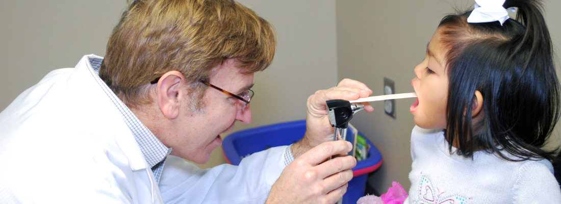
During an examination, your ENT specialist will use an otoscope to look inside and assess your ear. They check for redness in the ear, and/or fluid behind the eardrum, and to see if the eardrum moves. These are the signs of an ear infection. If your hearing is decreased, your ENT specialist may also perform an audiogram to test for any potential hearing loss by presenting tones at various pitches, or a tympanogram, which measures the air pressure in your middle ear to see how well your eustachian tube is working.
Your ENT specialist may also prescribe medications, which must be taken as directed. Often, antibiotics to fight the infection will make your earache go away rapidly, but the infection may need more time to clear up. Other medications that your doctor may prescribe include an antihistamine (for allergies), a decongestant (especially with a cold), or both. Sometimes the doctor may recommend a medication to reduce fever and/or pain. Special ear drops can also help ease the pain.
Children who experience multiple episodes of acute otitis media within a short time, chronic otitis media that lasts for more than three months, and/or hearing loss may require the insertion of ventilation tubes, also called pressure-equalization (PE) tubes. This is a short surgical procedure in which a small incision is made in the eardrum, any fluid is suctioned out, and a tube is placed in the eardrum. This tube will eventually fall out on its own, and the eardrum heals.
Remember, without proper treatment, damage from an ear infection can cause chronic or permanent hearing loss as well as more severe infections to the surrounding important structures.
What Questions Should I Ask My Doctor?
- How can I avoid earaches in the future?
- How do I clean my ears?
- Can my eustachian tube function be improved?
- Are ear tubes recommended for preventing ear infections?
Dysphagia
Dysphagia means that you can’t swallow well. Dysphagia is not a diagnosis; it is the symptom. Many factors may cause dysphagia, and most are temporary and non-life-threatening. In uncommon situations, swallowing difficulties can be related to a tumor or a nerve system disorder. It happens to people of all ages, but more often in the elderly. If swallowing is difficult on a regular basis, you should see an ENT (ear, nose, and throat) specialist, or otolaryngologist.
People normally swallow hundreds of times a day to eat solids, drink liquids, and swallow the normal saliva and mucus that the body produces. The process of swallowing has four related stages:
- The first stage is the oral preparation stage, where food or liquid is made ready in the mouth, chewed, and gathered together in preparation for swallowing.
- The second stage is the oral stage, where the tongue pushes the food or liquid to the back of the mouth, starting the swallowing response.
- The third stage is the pharyngeal stage, when what is processed in the mouth is passed through the pharynx, your throat, and into the esophagus, your food pipe.
- In the fourth and final stage, the esophageal stage, food or liquid passes through the esophagus and into your stomach.
The third and fourth parts of the swallowing process happen automatically, without you even thinking about it.
What Are the Symptoms of Dysphagia?
Symptoms of swallowing disorders may include:
- Drooling
- A feeling that food, liquid, or pills are sticking in the throat
- Coughing or choking on bits of food or liquid, or saliva not moving easily, which may lead to aspiration (when these materials fall or get sucked into the lungs)
- Sensing of a “lump” in the throat
- Losing weight
- Developing lung infections like pneumonia
- Changing voice
- Coughing up blood
What Causes Dysphagia?
Dysphagia may result from one or more of these issues:
- Acid reflux
- Throat infections (such as tonsillitis)
- Age-related swallowing muscle weakness
- Food or other foreign body becoming stuck in the throat (particularly in older patients)
- Weakness or scar of the esophagus
- Vocal fold paralysis or weakness
- Side effect of medications
- Tumors (throat, lung, esophageal cancer)
- Prolonged illness needing long stays at the hospital
- Past surgery or radiation to the neck, back, or chest
- Nerve disease such as Parkinson’s disease, multiple sclerosis (MS), amyotrophic lateral sclerosis (ALS, also known as Lou Gehrig’s disease), Myasthenia Gravis, or stroke
What Are the Treatment Options?
Your ENT specialist may work with other healthcare specialists, such as a gastroenterologist (GI), neurologist, and/or speech-language pathologist (SLP), to accurately diagnose and effectively treat the source of the problem.
When dysphagia is frequent, and the cause is not clear, your ENT specialist will discuss the history of your problem and examine your mouth and throat. They may insert a small tube called a flexible laryngoscope through your nose to help them examine your throat in greater detail. Sometimes, giving you food or liquid while the scope is in place helps them get a better look at the back of your tongue, throat, and voice box (larynx), and see what happens when you swallow. This procedure is called Flexible Endoscopic Evaluation of Swallowing (FEES).
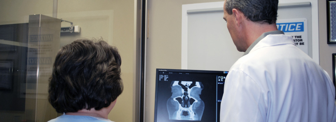
Your doctor and/or specialist may also order other tests like the barium swallow (or esophagram) and modified barium swallow. In these tests, instead of a flexible laryngoscope, X-rays record how food and drinks go down, and help your doctor evaluate the entire swallowing process. If necessary, they may do an examination of the esophagus, called Trans-Nasal Esophagoscopy (TNE), or refer you to a GI doctor for an upper endoscopy, which evaluates the esophagus and stomach with a flexible camera. TNE is similar to flexible laryngoscopy except the scope is longer and is passed all the way to the stomach. Additional testing can include pressure testing (manometry), which evaluates pressure created by the throat and esophagus muscles to see if they are working correctly.
If you have trouble swallowing, it is important to seek treatment to help you avoid malnutrition, dehydration, and pneumonia.
What Questions Should I Ask My Doctor?
- Do you know why I have difficulty swallowing?
- What are the tests for my swallowing problem?
- Can anxiety cause swallowing difficulty?
- What is the treatment for dysphagia?
- What is swallowing therapy?
- Do I need an endoscopy?
- How do you decide if I need a feeding tube?
- If I need a feeding tube is it permanent?
Deviated Septum
The bone and cartilage that divides the inside of the nose in half is called the nasal septum. The bone and cartilage are covered by a special skin called a mucous membrane that has many blood vessels in it. Ideally, the left and right nasal passageways are equal in size. However, it is estimated that as many as 80 percent of people have a nasal septum that is off-center. This is called a deviated septum, which may or may not cause certain symptoms.
What Are the Symptoms of a Deviated Septum?
The most common symptom from a badly deviated or crooked septum is difficulty breathing through the nose, which is usually worse on one side. In some cases, a crooked septum can interfere with sinus drainage and cause repeated sinus infections. You may experience one or more of the following:
- Difficulty breathing through one or both nostrils
- Nosebleeds
- Sinus infections
- Noisy breathing during sleep in infants and young children
- Mouth-breathing during sleep in adults
What Causes a Deviated Septum?
Injury or trauma to the nose can cause the septum to become deviated or crooked. However, even people with normal growth and development, and without a history of injury, trauma, or broken nose, can have a deviated septum.
What Are the Treatment Options?
Discuss your symptoms and any known nose damage or surgeries with your primary care provider or an ENT (ear, nose, and throat) specialist, or otolaryngologist. They will examine your nose inside and out, and might recommend additional tests based on your individual needs. When there is clearly a crooked/deviated septum, and the symptoms are severe enough to warrant intervention, the ENT specialist may suggest surgery as an option if medical treatment fails.
Septoplasty is the preferred surgical treatment to correct a deviated septum. This procedure is typically not performed on young children, unless the problem is severe, because facial growth and development are still occurring. Septoplasty is a surgical procedure that is usually performed through the nostrils, so there is no bruising or outward sign of surgery; however, each case is different and special techniques may be required depending on the individual patient.
The time required for the septoplasty operation averages about one- to one-and-a-half hours, depending on the type of deformity. It can be done with a local or a general anesthetic, usually on an outpatient basis. During the surgery, badly deviated portions of the septum may be removed entirely, or they may be readjusted and reinserted into the nose. Surgery may be combined with a rhinoplasty that changes the outward shape of the nose; in this case swelling and bruising may occur. Septoplasty may also be combined with sinus surgery.
Are There Related Factors or Conditions?
- Inferior turbinate hypertrophy—turbinates are finger-like structures in your nose that warm and moisten the air you breathe, and sometimes the lower ones can get too big
- Concha bullosa of the middle turbinate—this is when one of the turbinates next to your sinus openings gets a big air bubble in it
- Nasal valve collapse (internal or external)
- Sinusitis (acute, recurrent, chronic)
- Headaches (contact point)
- External nasal deformity (change in the shape of the nose)
- Decreased sense of smell
Are There Potential Dangers or Complications?
Sometimes a deviated septum may lead to repeated nose bleed . If the blockage is severe, it may force mouth-breathing at night, which can worsen sleep disorders. However, potential complications from septoplasty (surgery) can include:
- Anesthesia complications
- Bleeding
- Infection
- Creation of a hole connecting the right and left sides of the nasal cavity (called a septal perforation)
- Numbness of the upper teeth and nose
- Cerebrospinal fluid leak (extremely rare)
- Change in the external shape of the nose
What Questions Should I Ask My Doctor?
- I’ve been experiencing some of these symptoms. Is it possible that I have a deviated septum and, if so, how severe do you think it is?
- Is there anything about my nose that might be interfering with my breathing?
- Are there any other conditions that may be contributing to my nasal congestion/obstruction?
Cricopharyngeal Muscle Dysfunction
If the cricopharyngeal muscle (CPM) in your throat malfunctions or is impaired, this can cause you to have difficulty swallowing. The top valve of your esophagus (food pipe) is called the upper esophageal sphincter (UES), or pharyngoesophageal segment (PES). The CPM separates the esophagus and throat. Unlike most muscles, the CPM remains flexed and tightly closed unless nerves signal it to relax. This protects the throat and windpipe from food or liquid coming back up and inadvertently entering the lungs. For food, liquid, and saliva to enter the esophagus, the CPM needs to relax while these contents pass through into the esophagus.
What Are the Symptoms of CPM Dysfunction?
If you have CPM dysfunction, you may experience:
- Sensation of food sticking in the back of the throat
- Increased effort when swallowing food, liquids, saliva, pills, etc.
- Coughing when eating or drinking
- Choking episodes
- Inability to swallow large pieces of food
- A “wet” sounding voice
- Aspiration pneumonia
- Fear of eating
What Causes CPM Dysfunction?
CPM and UES dysfunction occur for varying reasons. It may result as a side effect of the normal aging process or due to changes in the CPM or nerve signaling pathways. Specifically, muscle enlargement (called hypertrophy), scarring of the muscle (called fibrosis) from radiation therapy or trauma, stroke, and reflux (heartburn) can all damage the swallowing mechanism at the UES.
To test for CPM and/or UES dysfunction, your ENT (ear, nose, and throat) specialist, or otolaryngologist, may examine your throat and larynx (voice box) by passing a small flexible camera through your nose. In addition, your ENT specialist may order an X-ray swallowing test called a modified barium swallow, esophagram, and/or manometry (pressure testing) of the valve area.
During the modified barium swallow, also known as a Videofluoroscopic Swallow Study (VFSS), you swallow various barium-coated food, liquid, and pills while a speech language pathologist takes X-ray video. For an esophagram, a radiologist looks at the esophagus with X-rays as barium is swallowed in order to evaluate esophageal function.
In CPM dysfunction, a narrowing of the valve may be seen, although narrowing of the PES may not be abnormal or require intervention. Manometry is a test that measures throat and esophageal pressures during swallowing by placing a thin tube through the nose into the esophagus and having patients swallow water. In CPM dysfunction, the valve may have high pressures at rest and/or during swallows.
What Are the Treatment Options?
There are numerous treatment options that can substantially improve CPM dysfunction, including dilatation (stretching), oral medications, BOTOX® injection, and myotomy (cutting). Dilatation procedures are generally not permanent and allow for a trial period to see whether a treatment will be helpful. These can be done either with mild or no sedation, or under general anesthesia.
As with stretching, BOTOX injections into the CPM provide temporary results, generally for three to six months, by causing partial paralysis or weakening of the CPM. This procedure is most often done in the operating room. Cricopharyngeal myotomy, or cutting the CPM with a laser or surgical instrument, can be done either endoscopically (through the mouth without any incisions on the neck) or through the neck skin, depending on the patient.
Treatment of CPM dysfunction can provide significant improvement in swallowing and quality of life. Duration of improvement often depends on the underlying cause of the dysfunction. It is not uncommon for patients with good outcomes after stretching to proceed to CPM myotomy for a permanent solution if you and your doctor decide this is the best course of action for you. Overall, most patients do well with procedures, and experience few side effects or complications.
What Questions Should I Ask My Doctor?
- Why do I have trouble swallowing?
- What tests can help determine why I have a swallowing problem?
- How do I know if my swallowing problem is related to my stomach, my esophagus, or my throat?
- Does my swallowing problem put me at risk for aspiration?
- Do I need to have further testing for my swallowing difficulty?
- What if I do nothing for my swallowing problem; can it be managed without a procedure?